Cattle Information

Information on common bovine problems

Mastitis

Diagnosis of Mastitis in Cows

Early identification of all cases of clinical mastitis is a key weapon in preventing further outbreaks of mastitis and is a key part of the overall mastitis control programme, ensuring maximum herd health, milk quality and dairy farm profitability.

The organisms that cause mastitis in dairy cows are divided into 2 main groups; contagious and environmental mastitis. There is some degree of interplay between these two types, for example strep uberis is primarily an environmental organism that can become ‘cow adapted’ and change into a contagious organism.

Each type of mastitis may present in an entirely different way. For example as infection with Staphylococcus aureus progresses through a herd the incidence of clinical mastitis may be quite low but the herd cell count continues to rise.

Streptococcus uberis may present with a high level of clinical cases and fluctuating bactoscans and herd somatic cell counts. Once ‘cow adapted’ strains of Strep uberis develop the herd cell count will increase rapidly due to the contagious transfer of infection.

Each farm has its own mastitis profile and the first task when investigating a problem is to establish the main organisms involved.

This can be assessed in two ways:

Take a bulk milk sample under strict conditions and submit it for specialist examination.

Take samples from each case of mastitis and store them in the freezer. These can then be analysed at a later date for the causative organisms.

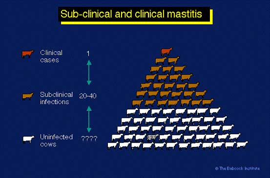
Subclinical Mastitis

The most important economic loss from mastitis is subclinical mastitis, for every clinical case of mastitis in a dairy herd there can be 25 or more subclinical cases. These subclinical cases are the main cause of increased cell counts:

https://WEU-AZ-WEB-CDNEP.azureedge.net/mediacontainer/medialibraries/threeriversvetgroup/images/general/subclinical-clinical-mastit.jpg

Mastitis Risk Factors - Five Point Plan

The main risk factors for mastitis are shown below:
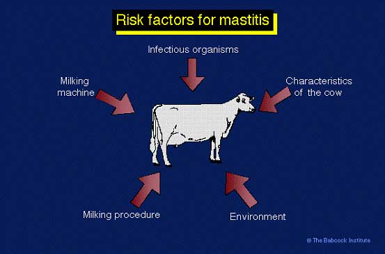
https://WEU-AZ-WEB-CDNEP.azureedge.net/mediacontainer/medialibraries/threeriversvetgroup/images/general/risk-factors-mastitis-cows.jpg

The Five Point Plan

The five point plan was established by the Milk Marketing Board in the 1950’s in order to counteract the mastitis risk factors – the points are still valid today:

  • Hygiene: i.e. improving operator hygiene during milking such as the wearing of gloves and using disposable teat wipes.
  • The correct maintenance of milking machine equipment.
  • Post-milking teat disinfection.
  • The prompt identification of clinical cases, veterinary treatment and the culling of chronic recurrent cases.
  • Dry Cow Therapy.

Contagious Mastitis

The main contagious organisms are listed below:

  • Staphylococcus aureus
  • ‘Cow-adapted’ Streptococcus uberis
  • Coagulase negative staph (CNS)

Other organisms which are quite rare but cause serious outbreaks of contagious mastitis are:

  • Streptococcus agalactia
  • Mycoplasma
  • Control of contagious mastitis

Staphylococcus aureus (S. aureus)

This bacterium is extremely difficult to control by treatment alone, because the response to antibiotic treatment is poor. Successful control is achieved by prevention of new infections and culling infected cows. S. aureus organisms colonize damaged teat ends or teat lesions. The infection is spread form cow to cow by a number of factors e.g. milkers’ hands, wash cloths, teat cup liners, and flies. The organisms probably penetrate the teat canal during milking. Irregular vacuum fluctuations impact milk droplets and bacteria against the teat end with sufficient force to cause teat canal penetration and possible development of new infection. Infected cows must either be culled, segregated from the milking herd and milked last or milked with separate milking units, or teat cup liners must be rinsed and sanitized after milking infected cows. The development of automatic dipping and flushing through the cluster has the potential to revolutionise the control of S. aureus mastitis.

Staphylococcus aureus causes chronic mastitis, often it is subclinical, where there is neither abnormal milk nor detectable change in the udder, but somatic cell count has increased. Some cows may flare-up with clinical mastitis, especially after calving. The bacteria persist in mammary glands, teat canals, and teat lesions of infected cows and are considered contagious. The infection is spread at milking time, when S. aureus contaminated milk from infected cows comes into contact with teats of uninfected cows, and the bacteria penetrate the teat canal. Once established, S. aureus often does not respond to antibiotic treatment, and infected cows eventually must be segregated or culled from the herd. Eradication of S. aureus, has proved to be almost impossible; however control is possible by practicing the highest level of hygiene and milking techniques.

Cows infected with S.aureus do not necessarily have high SCCs. Only 60% of infections are found in cows producing milk with more than 200,000 SCC. In several research trials, 3-8% of first lactation cows were found infected with S. aureus at calving. Many remain infected throughout the first lactation and are reservoirs for infecting other cows in the herd. Although as many as half of the cows with high SCC may be infected with S. aureus, somatic cell counts alone are not sensitive enough to positively diagnose S. aureus infections.

Streptococcus agalactiae

This infection is now quite rare; it is caused by poor hygiene and milking plant maintenance. Take care not to buy it in with replacements!

Since 2000 there has been a small rise in incidence due to corner cutting as a result of poor milk prices.

The infection causes massive bacterial counts 10 million or more in a cow!

The organism inhabits ducts and cisterns in the mammary gland, however it does not survive in environment and can be eliminated by simultaneous herd treatment.

The infection causes inflammation which blocks ducts and leads to decreased milk production and increased somatic cell counts.

Control of Contagious Mastitis

Quarantine Infected Cows

Keep infected cows in a separate treatment group and milk them last or use a separate claw and bucket.

Teat Dipping - Germicidal Dips

Postmilking teat dipping with germicidal dips is recommended. Barrier dips are useful against environmental infection but their effect against the contagious pathogens appears to be lower than that of germicidal dips.

Dry Cow Therapy

Treat all quarters with dry cow antibiotics at end of lactation, this is essential.

Special Treatments at Drying off

Some cows with a history of mastitis or raised cell counts require special additional treatment t drying off.

ADF (auto dip/fush) - (backflushing)

The cost of these units is coming down all the time; probably the best way of stopping transfer of infection.

Flush milk claws (hot water or germicide) after milking infected cows

If using a germicide ensure that it is licenced for this purpose.

Use individual cloth/paper towels to wash/dry teats

Hygiene

Clean hands (wear gloves). good hygiene and teat preparation.

Milking Machine Maintainence

Malfunctioning milking machines that result in frequent liner slips and teat impacts can increase cases of environmental mastitis. Ensure your machine is cheched by a technician regularly and any faults rectified as soon as possible. Also make sure your liners are replaced at the correct time – a simple calculation is shown below:

Liner life = 2,500 x number of milking units
Number of cows x 2*
* milked twice daily.

A poorly maintained milking plant will cause teat end lesions which allow infections to enter the teat canal.

Avoid buying infected cows

Always check cell count history of individual cows and the herd of origin as a minimum requirement.

Cull persistently infected cows

Environmental Mastitis

The main causes of environmental mastitis are:

  • Escherichia coli (E.Coli)
  • Streptococcus uberis
  • Pseudomonas species

Streptococcus uberis (Strept uberis)

Strepuberis mastitis is probably the most important organism with regard to mastitis in the modern dairy cow. It is an environmental organism (Control of environmental mastitis) that lives in faeces, the rumen and in all areas where there is cow contact. Whilst it is an easy organism to kill in a petri dish it is very difficult to eradicate from an infected udder. The organism may progress from being an environmental cause of infection to a contagious cause by becoming cow-adapted (Control of contagious mastitis). In other words the organism lives in the udder and transmits to other cows during the milking process in the same way as S.aureus. Large clinical outbreaks of strep uberis can result in high and fluctuating bactoscans as well as raised herd SCC.

Treatment of Strep uberis mastitis must be aggressive and targeted with specific treatment protocols. If treatment is not aggressive then there is more likelihood of cow-adapted infections developing. This is why many farms fail in their attempts to control and treat strep uberis mastitis.

Pseudomonas

The clinical signs are similar to E. coli. Pseudomonas mastitis is usually a result of a hygiene problem in the parlour. It generally causes infection from contaminated water, pipes, heater, wash hoses, teat dip. It is often resistant to many antibiotics

Control of Environmental Mastitis

1. Housing and Environment

Housed cows are at greater risk for environmental mastitis than cows on pasture. However in the summer oubreaks of environmental mastitis often occur, these are due to either high rainfall or very hot weather which causes the cows to shelter under trees and produce a very contaminated area. Sources of environmental pathogens include manure, bedding, feedstuffs, dust, dirt, mud, and water.

Bedding materials are a significant source of teat end exposure to environmental pathogens. The number of bacteria in bedding fluctuates depending on contamination (and, therefore, availability of nutrients), available moisture, and temperature. Low-moisture inorganic materials, such as sand or crushed limestone, are preferable to finely chopped organic materials. In general, drier bedding materials are associated with lower numbers of pathogens. Warmer environmental temperatures favor growth of pathogens; lower temperatures tend to reduce growth.

Finely chopped organic bedding materials, such as sawdust, shavings, recycled manure, pelleted corncobs, peanut hulls, and chopped straw, frequently contain very high coliform and streptococcal numbers. With clean, long straw, coliform numbers are generally low; but the environmental streptococcal numbers may be high.

Attempts to maintain low coliform numbers by applying chemical disinfectants or lime are generally impractical because frequent, if not daily, application is required to achieve results. Total daily replacement of organic bedding in the back third of stalls has been shown to reduce exposure of teat ends to coliform bacteria.

Environmental conditions that can increase exposure include: overcrowding; poor ventilation; inadequate manure removal from the back of stalls, alleyways, feeding areas and exercise lots; poorly maintained (hollowed out) free stalls; access to farm ponds or muddy exercise lots; dirty maternity stalls or calving areas; and general lack of farm cleanliness and sanitation.

Control of environmental mastitis is achieved by decreasing teat end exposure to potential pathogens or by increasing the cow’s resistance to mastitis pathogens.

2. Teat Dipping - Germicidal Dips

Premilking dipping is advocated for the control of environmental pathogens and is certainly recommended in herds where there is a high risk of either E.coli or Strep uberius infections.

Postmilking teat dipping with germicidal dips is recommended. A degree of control over the environmental streptococci is exerted, but there is no control of coliform intramammary infection.

3. Teat Dipping - Barrier Dips

Barrier dips, postmilking, are reported to reduce new coliform intramammary infections. Their efficacy against the environmental streptococci and the contagious pathogens appears to be lower than that of germicidal dips.

4. Dry cow therapy and teat sealants

Dry cow therapy with the use of teat sealants of all quarters of all cows is recommended. Dry cow antibiotic therapy significantly reduces any infections carried over from the previous lactation and the teat sealant prevents new environmental infections.

5. Backflushing or use of germicidal sprays between cows

Ensure the product you are using is licenced to be used in this way. This proceedure is usefull in preventing the spread of cow-adapted strep uberis infection, However it does not prevent initial environmental mastitis infection.

6. Milking machine problems

Malfunctioning milking machines that result in frequent liner slips and teat impacts can increase cases of environmental mastitis. Ensure your machine is cheched by a technician regularly and any faults rectified as soon as possible. Also make sure your liners are replaced at the correct time – a simple calculation is shown below:

Liner life = 2,500 x number of milking units
Number of cows x 2*
* milked twice daily.
A poorly maintained milking plant will cause teat end lesions which allow infections to enter the teat canal.

7. Udder preparation

Milking cows with wet udders and teats is likely to increase the incidence of environmental mastitis. Teats should be clean and dry prior to attaching the milking unit. Washing the teats, not the udder, is recommended.

8. Vaccination

There is a vaccine available for mmunizing cows during the dry period against Escherichia coli J-5 bacterin. This vaccine will reduce the number and severity of E.coli clinical mastitis cases during early lactation.

9. Diet

Feeding diets deficient in vitamins A or E, beta-carotene, or the trace minerals selenium, copper, and zinc will result in an increased incidence of environmental mastitis. The supplementation of the dry cow diet with Selenium has been shown to be of benefit.

Hoof Health

Lameness in Dairy Cows

After infertility and mastitis, lameness is the third most common reason for culling cows on dairy farms.  In the average dairy herd 38 out of every 100 cows is affected by lameness per year, some cows being affected more than once.  90% of lameness cases involve the feet and 90% of these involve the rear feet, the majority affect the outside claw. Lameness prevention is much better than treatment from both an animal welfare and an economic point of view. Locomotion scoring can be used to asses the progress of a lameness action plan, which is likely to include regular foot bathing and foot trimming.

This costs the average 100-cow dairy herd almost £4,595 per year (i.e. approximately 1pence per litre of milk produced) these losses are a result of:

  • Reduced milk yield
  • Increased calving interval
  • Increased inseminations per pregnancy
  • Increased culling rate
  • Cost of treatments, inc. herdsman’s time

Types of lameness:

  • Digital dermatitis.
  • Laminitis.
  • Sole ulcer.
  • White line disease.
  • Slurry heel erosion.
  • Foul of the foot.
  • Toe necrosis.
  • Horizontal vertical wall fissures.
  • Hock lesions.
  • Trauma, injury and foreign body penetration of the sole.

Lameness Prevention

Any lameness action plan must take into account the factors below if it is to be successful.

1. Environmental Factors

  • Enable the cow to enter and leave cubicles easily
  • Ensure the cow can lie down and rise without interference
  • Ensure the cow can stand or lie in the stall comfortably
  • Avoid the rear feet constantly being in contact with slurry and urine
  • Take care with slurry cleaning and scraping
  • Provide soft dry bedding – particularly for pre-calving heifers
  • Check flooring condition –? renew poor concrete, concrete grooving, avoid variable heights
  • Observe cows daily – watch for foot injuries
  • Treat cows promptly and place in straw yard/ pen for recovery
  • Regular foot bathing greatly reduces lameness in most cases
  • Regular foot trimming is also important

2. Nutritional Factors

  • Avoid Rumen acidosis
  • Take care in heifer rearing to avoid laminitis

3. Inherited Factors

  • Avoid selecting replacement heifers from cows with poor conformation, gait or hoof type
  • Use Bulls with good classification for leg and hoof conformation
  • Locomotion scoring can be used to assess the progress of a lameness action plan.

Mobility Scoring 

We have a Registered RoMS mobility scorer that can provide on farm services to our clients. The RoMS is an independent, self-regulatory body which encourages the widespread use of standardised, independent mobility scoring conducted by trained and accredited scorers on UK dairy farms. AHDB have developed a method for mobility scoring which is the industry standard in the UK and is the only methodology than can be used by members of the RoMS. 

Foot Bathing

Footbathing is an important part of the management and control of lameness – particularly digital dermatitis and slurry heel. Foot baths are best situated near the parlour exit – far enough not to reduce flow of cows.

Two baths are ideally required, the first for washing the feet and the second to apply the treatment. Foot baths may be concrete or plastic, they should be easy to set-up and clean out and near water and drainage. Some authorities recommend straw in first bath to produce better cleaning. If there is only one bath available then the feet should be power washed in the parlour before the cows leave.

Foot bath requirements:

  • Min. length 3 m, depth 20 cm
  • Flush to wall or rails of race
  • Non-slip floor, + ridges along long axis?
  • Standing room beyond bath

It is important to stick to a routine and locomotion score regularly.

The most common ingredients used in cattle footbaths contain one or more of the following chemicals:

1. Copper sulphate, 2.5 to 12.5

  • Proven efficacy
  • Can be expensive
  • Easy to use
  • Disposal is a serious environmental concern

2. New copper sulphate delivery system. Healthy Hooves

  • Reduces the copper sulphate used by 70%
  • Reduces the cost
  • More effective control of Digital Dermatitis
  • See our fact sheet (this links to fact sheet attached)
  • Link to Healthy hooves Web site http://www.healthyhooves.eu
  • Three Rivers Veterinary Group are stockists for Healthy Hooves products

3. Antibiotics

  • Expensive and not recommended from a farm assurance point of view
  • Only used in extreme circumstances
  • Withdrawal period issues

4. Organic acids

  • Have an antibacterial effect
  • Can be used with copper sulphate to prevent binding of copper sulphate by organic material on the foot

5. Zinc sulphate

  • Usually used in combination with copper
  • Good for healthy hoof horn

Foot Trimming

This is a vital part of a lameness control program.

For routine herd foot trimming you will need the services of a qualified foot trimmer. However for individual cases that occur between the foot trimmer’s visits you will need to treat them yourself or ask for our help.

We are happy to give individual tuition on foot trimming and corrective paring of diseased hooves, please contact us.

The following reference is a concise guide to foot trimming including the Dutch method, please log on to it.

Digital dermatitis appeared in the UK around 1987 and has since become one of the most serious causes of lameness (>25%), 90% of cases occur on the hind feet.

Digital dermatitis is caused by 3-5 different spirochaete bacteria (Treponema spp.) and it is most common on the underside of the hind foot above the heel. The infection causes circumscribed, eroded, painful lesions surrounded by hyperkeratosis and hypertrophied hairs. In long standing cases these hairs can become very marked and are known as ‘hairy warts’.

Affected cows are lame and slow and often stand and constantly lift the foot as is the pain appears to be sharp and stinging. Slurry heel or heel/horn erosion often progresses from the initial lesion.

The spirocheates thrive in wet conditions especially slurry; it is likely that the irritant effect of urine in slurry allows the organism to attack the skin. Cattle from affected farms carry the infection and most farms became infected due to the purchase of infected cows. Contaminated foot trimming equipment can also spread the disease.

The disease is controlled on a herd scale by foot bathing. Individual treatment may be required in more severe cases; we treat these with our ‘digital dermatitis cream’ and bandage plus in some cases antibiotic treatment.

The cost of each case of digital dermatitis is approx £106 Young, S., MDC Study. An average 38% incidence in a 100 cow herd gives a total annual cost of around £4,028.

Laminitis

Laminitis is the route cause of many herd lameness problems and it’s control can make spectacular savings to the annual lameness treatment costs.

There are three types of laminitis:

1. Acute Laminitis

  • Sudden onset
  • Severe pain
  • All four legs affected
  • Difficulty in standing – or cow will kneel only
  • Not common

2. Sub-acute Laminitis

  • Most common form of laminitis on farm
  • No obvious signs
  • Frequent nutritional and environmental stress
  • Heifers on high cereal/silage diets
  • Cows on hard standing for long periods
  • Hoof weakened through abnormal growth
  • Yellow sole, haemorrhaging and sole ulcers

The following are associated with sub-acute laminitis:

  • Sole ulcer
  • White-Line Disease
  • Toe necrosis
  • Double or false sole

3. Chronic Laminitis

  • If left untreated, sub-acute laminitis will progress to chronic laminitis
  • Breakdown of blood supply to the corium and damage to the internal structure of the foot
  • Irreversible damage, culling

The link between nutrition and laminitis is acidosis which results in poor blood supply to the corium. The severity depends upon frequency and length of each period of rumen acidosis.

Sole Ulcer

Sole ulcers are caused by the uneven distribution of weight in the foot. The increased pressure on the inside of the sole causes damage and necrosis of the tissues below the horn and an ulcer develops. Sole ulcers are preventable by regular foot trimming and hoof care; they are a major cause of economic loss. Often the opposite claw requires a block glued on to it to take the weight off the affected claw and allow it to heal. The healing process takes some considerable time and there is always a weakness there afterwards.

Sole ulcers are one of the 3 most common causes of lameness; the others are digital dermatitis and white-line disease. Sole ulcers cause the most-severe lameness and they are also expensive the average cost of a case is £314.08 (Esselmont).

Regular foot bathing helps to improve horn quality and reduce the incidence of sole ulcers.

White Line Disease

The white line lies between the hard side wall of the hoof and the more pliable sole. It allows some elasticity and mobility within the hoof and the line is part of the lamellae which lock the hoof onto the pedal bone. Reduced horn quality and hardness leaves the structure more susceptible to damage and vascular disturbances. It is known that when the white line is weakened foreign material (grit) may be caught in it and gradually work its way into the foot with the pressure and shearing forces as the cow moves along especially on hard surfaces. If foreign material ascends the white line and penetrates the sensitive tissue of corium, infection will most likely occur, which leads to lameness and further damage to the white line at the point of origin.

A regular hoof trimming program and attention to the hoof quality with regular foot bathing of the herd will assist in spotting potential white line disease. The elimination of standing water and maintenance of high quality walkways will help to ensure healthy hoof horn structure. Old damaged concrete surfaces are a source of hard grit and this often causes white line disease.

Slurry/Heel Erosion

Slurry heel or heel erosion is associated with interdigital dermatitis. The cause is multifactorial with a bacterial component superficial. The skin becomes greyish, sweaty, smelly with no obvious lameness. The inflammation extends to the heel and erodes it, V-shaped fissures appear and poor-quality black horn is produced.

There is a reduction of horn production at the heel and an increase in horn production elsewhere in the hoof: lengthening, sometimes with elevation. Regular foot bathing helps to improve horn quality and reduce the incidence of slurry/heel erosion.

Mild lameness may appear and various complications arise such as:

  • Double or false sole
  • Bruised sole
  • Sole ulcer

Foul of the Foot

Foul of the foot is a contagious disease of cattle characterized by the development of a necrotic lesion in the interdigital skin. The accompanying infection extends into the soft tissues of the foot causing swelling and lameness. The lesion has a typically foul smell which some find helpful in distinguishing it from other conditions.Incidence appears to be higher during the winter months and in confinement-housed cattle.

Recently a new more severe form of the disease has been observed. It has been termed “Super Foul” which is believed to result from a combined infection with the spirochaetes that cause digital dermatitis; very prompt aggressive treatment is required in these cases if the cow is to be saved.

Environmental factors may cause foul of the foot such as walking through or standing in manure slurry for extended periods or flints and stones in gateways or walkways. The condition is caused by Fusobacterium necrophorus and it lives in the gastrointestinal tract of the cow.

Improving the quality of walkways and alleyways and eliminating debris in these areas to avoid interdigital skin lesions and keeping the cattle environment as dry as possible are major considerations in the prevention of foot rot problems.

Treatment requires systemic antibiotic therapy and simple cases will usually respond to penicillin alone.

Regular foot bathing helps to control the infection reduce the incidence of foul of the foot.

Toe Necrosis

This is a serious infection of the toe with the spirochaete organisms that cause digital dermatitis. The infection has a gangrenous smell and gradually eats away the toe; initially the cow may not be too lame. Regular paring can ease the problem in the early stages but as the disease progresses the animal will have to be culled.

Horizontal and Vertical Wall Fissures

Horizontal fissures in the outside wall of the hoof are usually a result of a severe illness or metabolic disorder which affected hoof production from the coronary bane for a period of time. These fissures will grow out but they can be painful at times, corrective paring of the fissure to make it shallower often helps.

Horizontal fissures are often a result of either weak horn due to inherited or nutritional factors or they may be caused by a traumatic event e.g the foot becoming wedged somewhere.

Provided the horn has the capacity to regrow good quality, hard horn these defects will resolve themselves; however because of the slow growth rate of horn this takes a considerable time.

Hock Lesions

Hock lesions are associated with cubicle design problems. The type of bedding material also affects the incidence of hock lesions. Hock lesions and swellings may become infected and spread to the joint and it is difficult to save these cows.

On some farms poor cubicle design and maintenance may lead to a high number of cows with hock lesions and a herd welfare issue.

Fertility

Fertility Control Program

The Three Rivers Veterinary Group fertility control program covers the following areas:

  • Post calving period. All cows are checked post calving to ensure that there is no uterine infection, cystic ovaries or post calving damage which might affect fertility.
  • Oestrus detection target period of 42 days post calving. Any cows not observed in oestrus by day 42 post calving are examined and treated to get them cycling again.
  • Early detection of anoestrus cows with the use of target dates (i.e. those cows which cycle once post calving and then go quiet).
  • Early pregnancy and non pregnancy detection. We are able to perform early pregnancy diagnosis by ultrasound from day 28 and manual pregnancy diagnosis from day 35.
  • Monitoring of Body condition scores. This is routinely done in conjunction with VetImpress to provide feedback to the nutritionist regarding dietary balance.
  • Metabolic profiles taken at an early stage of lactation in conjunction with samples from cows about to calve and mid lactation cows provides valuable information about the state of the cows with regard to energy and protein balance and allows for alterations in the ration in time to affect fertility performance.
  • Regular monitoring of Interherd and Total Vet information in conjunction with the farm record to ensure a quick response to any problems.
  • Monitoring of bulk milk samples for disease e.g.:
  • Bovine Virus Diarrhoea (BVD)
  • Leptospira hardjo
  • Infectious Bovine Rhinotracheitis (IBR)
  • Liver Fluke
  • Gastro-intestinal worms

Bovine Embryo Collection and Transfer

Embryo transfer (ET) is a technique that can greatly increase the number of offspring a high merit cow can produce. An ET programme provides a faster genetic improvement than Artificial Insemination (AI) since both the female and male genetics are selected and amplified. This can help you to select healthier, more efficient animals which are better adapted to farm conditions and management, thus promoting welfare and reducing costs on your farm. 

Embryo transfer also minimizes the risks of transmission of most infectious diseases when embryo collection, donor, sire and recipient health testing and embryo freezing are conducted under OIE/IETS standards.

Embryo transfer requires two steps: generating and then obtaining (flushing) the embryos from the donor female and transferring each embryo into a different female (recipient) which gestates and give birth to that calf. These two components do not necessarily have to be done in the same farm. Embryos can be produced, frozen, sold and implanted in someone else cattle. So entire herds can be transported frozen in a liquid nitrogen tank at a cost that is often lower than for transporting a single animal.

Contact us for further information about our Embryo collection and transfer services.

BVD Stamp It Out

BVD Stamp It Out England

Bovine Viral Diarrhoea (BVD) is an endemic disease which affects cattle. The disease costs dairy and beef businesses an estimated £61 million/year through the loss of performance, infertility, mortalities and slower growth rates. DEFRA has made available £5.7 million of funding through the Rural Development Funding Programme in England (RDPE), aiming to raise the profile of the BVD virus in England. The project will engage veterinarians and farmers to investigate BVD at farm level, creating action plans to control, reduce and eliminate the disease. Opportunities will also arise to discuss matters with other likeminded farmers, discussing the most effective ways to manage the disease.

Contact us to apply to the programme.

Schmallenberg Virus

What is the Schmallenberg Virus?

The Schmallenberg virus is a newly emerging virus in the same family as the Akabane virus. Little is known about it, but new information is coming to light daily. As it is a new virus testing is still being developed and a vaccine is a long way off.

The virus appears to be spread by biting insects most likely midges (similar to bluetongue), however, direct transmission may also occur. Acording to Met office data there were 4-8 days in August to late October when infected midges could have been blown across the channel. Thus disease is emerging now but infection actually took place last year.

It appears that animals which are bitten by infected insects are highly likely to contract the disease, and subsequent insects biting this animal are highly likely to become infected themselves. Spread of the disease therefore is a lot quicker and easier than we have seen with bluetongue.

What does it do?

The clinical signs of Schmallenberg virus seem to primarily be seen in the foetus. These clinical signs consist of severe brain damage/deformities if infection occurs in the first third of pregnancy, limb deformities if infection occurs in the middle third of pregnancy, and brain problems presenting as depression, problems standing/balancing, and inability to suckle if infection occurs in the last third of pregnancy.

Infection in the animal usually has no symptoms at all, although in cattle it may present as diarrhoea, high temperature, and reduced milk yield.

Newborn animals that are affected may or may not be viable, whether the viable effected offspring are a disease risk to other animals is not known, it would be wise to separate any affected offspring with their dam, from the rest of the flock/herd.

At the present time infection has only been confirmed in sheep, but all ruminants are susceptible. Infection rates are variable, but up to 50% of offspring can be affected.

Can it infect humans?

Some viruses in the same family as this can infect humans but it seems Schmallenberg lacks the required genes. Having said this we would recommend women who are pregnant or hoping to become pregnant should not come into contact with ruminant animals, especially around lambing/calving/kidding.

What should I do?

At present the disease is not notifiable, but we encourage you to report any birth abnormalities to us at Three Rivers and Chapelfield Farm Vets.

The DEFRA website is updated daily with all the latest information, so this is a good place to start if you have questions or concerns.

Bluetongue Virus

Bluetongue Virus

In the autumn of 2006 Bluetongue (serotype 8) was found in the Netherlands, Belgium, Luxemburg, Western Germany and in parts of North Eastern France. The outbreak was identified as a new incursion in the EU. How Bluetongue entered Europe is still subject to investigation.

In August 2008 the first case of Bluetongue serotype 8 was diagnosed in East Anglia in a cow.

Bluetongue was first described in South Africa but has since been recognised in most countries in the tropics and sub-tropics. Since 1999 there have been widespread outbreaks of Bluetongue in Greece, Italy, Corsica (France) and the Balearic Islands (Spain). Cases also occurred in Europe in Bulgaria, Croatia, Macedonia, Kosovo and Yugoslavia. Serotypes 8, 1, 2, 4, 9 and 16 have been involved. These cases have been well north and west of its normal distribution. It appears that the virus has spread from both Turkey and North Africa. Bluetongue does not affect humans. A virus within the Orbivirus genus of the family Reorvirades causes bluetongue. At present 24 distinct serotypes have been identified as a result of serum neutralisation tests.

The virus is transmitted by a small number of species of biting midges of the genus Culicoides. Bluetongue virus cannot naturally be transmitted directly between animals. Virus transmission between animals occurs via the midges. However mechanical transmission of the virus between herds/flocks and within a herd/flock by unhygienic practices (e.g. use of contaminated hypodermic needles) is possible.

When a midge bites an infected animal, the virus passes to the midge in the blood meal and the virus multiples in the midge. The cycle of replication of the virus in the insect vector and in the ruminant host, results in amplification of the amount of virus available to naïve hosts and vectors. Peak populations of vector Culicoides occur in the late summer and autumn and therefore this is the time when Bluetongue is most commonly seen.

Clinical signs can vary by species – although symptoms are generally more severe in sheep, cattle are important in epidemiology of the bluetongue as they act as an often silent source of BTV – a reservoir for disease and keep the infection circulating.

There is a wide variation in the clinical signs seen in cattle

The initial signs in lactating cows are that the animal becomes slower and stiff with a reduced appetite and a fall in milk yield. There is usually a nasal discharge, clear at first with the mucosa inside the nostril noticeably redder than usual; this is accompanied by red eyes due to the inflammation of the conjunctivae. Over the following days erosions and crusting develop around the entrance to the nose; there may be crusty scabs on the teats. There may be some foamy saliva around the mouth. On opening the mouth there are erosive lesions on the upper gum (dental pad) which develop a yellowy skin of necrotic tissue over them, there may also be lesions on the hard palate and tongue. It is important to note that these are erosive lesions not vesicles as in Foot and Mouth! The lameness is due to a coronitis (i.e. inflammation of the coronary band which runs around the top of the hoof); if the foot is washed and cleaned the coronary band can be seen to be red and inflamed. Sometimes the leg above the coronary band may become secondarily infected and swell up.

There are problems with transplacental infection; pregnant cattle infected with Bluetongue will pass the infection on to their calf in utero. The calf may be aborted, die in the first few days of life or recover. It is believed that colostrum from recovered cows is valuable in helping weak calves to recover. These calves, their placentas and fluids will also act as a focus of infection on the farm, allowing the disease to flare up again in unvaccinated animals. The dams that have carried infected foetuses are likely to have lower milk production and fertility in their next lactation, reinforcing the point that prevention is the key in controlling this disease. If the disease spreads to a herd with a large number of pregnant animals the effects will be much more severe.

Bluetongue (BTV) update – APHA September 2018

BTV is a complex virus with 27 different strains, several of which are currently present in Europe: for example, BTV8 is present in France and Switzerland, BTV1 is present in France and BTV4 is present in Italy, Spain, Portugal and Cyprus (fig 8). In October, Italy reported nine outbreaks of BTV3 in Sardinia in sheep and goats. This is the first report of this serotype in Sardinia. BTV3 was first reported in the north of Tunisia in 2016, and then was identified in the north of Sicily in 2017. There is currently no inactivated vaccine available for this serotype. This reinforces the need to be aware of the different strains of BTV, what vaccines available and the specific requirements of each country if importation of animals is being considered. As the season progresses we should be aware that there may be a risk of wind-mediated midge transmission in at risk areas along the south coast of England but it may be difficult to estimate without knowing how heavy the infection pressure is in the North of France.

Following on from the detection in September of BTV through APHA post-import testing, two animals tested positive for BTV8 by PCR in a consignment of 60 steers imported from France. In October BTV was detected in a consignment of four sheep imported from France. One sheep was PCR positive for BTV-8 and all four were antibody negative to BTV suggesting that the vaccination which is required prior to export had not been effective or not been carried out. All four animals were euthanased. Surveillance is being undertaken in susceptible animals in the areas surrounding both of the UK destination farms at 4 and 10 weeks post-detection of BTV. Livestock owners have been reminded that they should source animals responsibly by working with their private veterinarians and livestock dealers to make sure animals are correctly vaccinated and protected prior to travel. This means that animals from France must be correctly vaccinated against BTV-4 and BTV-8 or be naturally immune to both virus serotypes, prior to leaving the Restriction Zone.

Bovine Respiratory Disease (BRD)

BRD - Pneumonia

The total UK calf population is 3.3 million and of these 30% get pneumonia (1million) each year; mortality varies from 3-50% (average ~5% or 157,000 calves a year).

Economic studies highlight the cost to national herd estimated of approximately £80 million; there are welfare costs too.

Three Rivers & Chapelfield Farm Vets pneumonia control program:

  • Reducing the risk of disease by quality advice regarding housing and environment solutions.
  • Good biosecurity and health planning.
  • Advice re vaccination programs.
  • Reduction of the necessity for on farm treatment
  • Reduction in antibiotic usage.
  • Achieving high quality farm assurance standards.

Environmental factors involved in calf pneumonia:

  • Poor ventilation
  • High humidity
  • Stress
  • Mixing age groups
  • Handling
  • Housing
  • Transport

The 'calf factors' in pneumonia incidence are:

  • Poor colostral intake
  • Nutritional status
  • Concurrent disease
  • Lung capacity

The viruses involved are:

  • Para-influenza 3 (PI)
  • Respiratory Syncytial virus (RSV)
  • Infectious Rhinotracheitis (IBR)
  • Bovine Viral Diarrhoea Virus (BVDV)

The bacteria involved are:

  • Pasturella multocida
  • Histophilus somnus
  • Mycoplasma Bovis

How do all these factors work together?

https://WEU-AZ-WEB-CDNEP.azureedge.net/mediacontainer/medialibraries/threeriversvetgroup/images/general/bvd.jpg

What are the common housing problems?

  • Lack of high level outlet for waste air.
  • Inadequate inlet areas for fresh air.
  • Shared airspace with older animals (who may carry the disease).
  • Low heat production from calves at low stocking levels.
  • High moisture levels (humidity above 70% is at a critical level for calves).
  • Poor design or location of mechanical ventilation.

Good ventilation is vital for the following reasons:

  • Removal of excess water vapour.
  • Removal of excess heat.
  • Removal of micro-organisms, dust and gases.
  • Provision of uniform distribution of air.
  • Provision of correct air speed for stock.
Return to Information for Farmers